Menu
Assessing Your Aorta Health
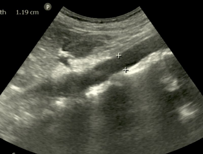
An abdominal aorta ultrasound scan is a specialized imaging procedure used to assess the abdominal aorta, the largest blood vessel in the abdomen. This non-invasive test employs high-frequency sound waves to create detailed images of the aorta, helping healthcare providers diagnose a variety of vascular conditions.


- Preparation: You may be asked to fast for several hours before the scan to ensure clearer images. It's best to wear loose, comfortable clothing.
- Procedure: During the ultrasound, you will lie on your back on an examination table. A gel will be applied to your abdomen to facilitate sound wave transmission. A transducer will then be moved over the area, capturing real-time images of the abdominal aorta and surrounding structures.
- Duration: The procedure typically takes about 20 minutes.
- Post-Scan: There are no side effects, and you can return to your normal activities immediately after the scan.
An abdominal aorta ultrasound scan is commonly performed to:
- Evaluate for abdominal aortic aneurysms (AAA)
- Assess the size and shape of the aorta
- Detect signs of atherosclerosis or other vascular diseases
- Investigate abdominal pain or other symptoms related to vascular issues
- Safe and Non-Invasive: This procedure does not use ionizing radiation, making it a safe option for patients of all ages.
- Real-Time Imaging: Allows for immediate visualization of blood flow and structural abnormalities.
- Quick Results: Images can be reviewed shortly after the scan, facilitating prompt diagnosis and management
Early Pregnancy Scan Package
- Confirmation of an early/viable pregnancy
- Measurement of the gestation sac and the Crown Rump Length (CRL)(if possible)
- Detection of heartbeat. (If Pregnancy is more than 6 Weeks).
- Between 6 and 10 weeks.
- 2x b/w images.
- 30 minutes appointment.